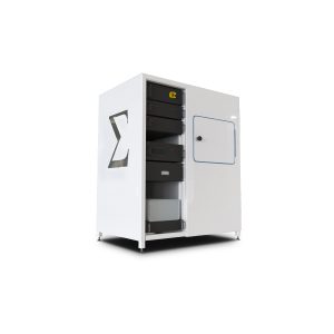
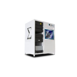
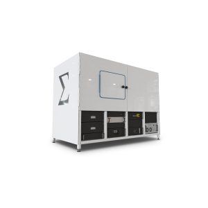
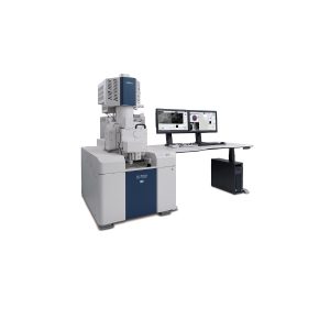
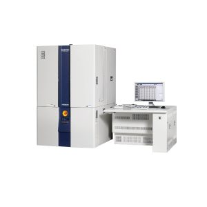
The Sigray EclipseXRM‑910 is a cutting-edge 3D X-ray microscope designed for laboratories that demand high-resolution imaging without relying on synchrotron facilities. With a 0.3 µm spatial resolution, it enables researchers to visualize submicron structures with exceptional clarity and precision.
Built for versatility, the EclipseXRM‑910 supports a wide range of samples, including materials, semiconductors, and biological specimens. Its advanced imaging capabilities allow scientists to explore internal structures, porosity, defects, and compositional variations in three dimensions, providing insights that are critical for research and quality control applications.
The system’s user-friendly software and intuitive workflow simplify experiment setup, scanning, and data analysis, reducing training time and improving laboratory efficiency. High-contrast imaging and flexible acquisition modes ensure reliable results for both routine characterization and complex research projects.
Compact yet powerful, the EclipseXRM‑910 fits easily into laboratory environments while delivering synchrotron-quality 3D imaging. Its combination of speed, precision, and versatility makes it ideal for academic research, industrial R&D, and advanced material studies.
With the Sigray EclipseXRM‑910, laboratories gain unlimited access to high-resolution X-ray microscopy, enabling faster insights, more accurate analysis, and accelerated discovery without the logistical constraints of shared facilities.

Unmatched Resolution and Efficiency for Superior Imaging Performance
The highest resolution achievable by the system is not only best-in-class, but is also achieved using a highly efficient detection system. This enables much higher throughput when imaging at smaller voxel sizes than other systems on the market.

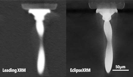
Superior Resolution and Speed in Imaging Carbonate Samples
EclipseXRM achieves higher resolution, faster: The same carbonate sample imaged on a leading XRM with two-stage magnification and EclipseXRM. The leading system advertises ~0.5 µm spatial resolution and used a 20X magnification objective to acquire the data in 4 hours, while EclipseXRM acquired 0.3 µm data in 2.5 hours.

Innovative Architecture for Submicron Resolution at Large Distances
EclipseXRM’s new, patent-pending architecture offers an alternative to prior approaches, enabling submicron resolution at large working distances without needing high magnification objective lenses.

Enhanced Contrast with Optional Multi-Spectral Source for Challenging Samples
SIGRAY EclipseXRM-910 optionally incorporates a Multi-Spectral Source, which produces quasi-monochromatic x-rays for outstanding contrast for challenging samples

Enhanced Contrast with Optional Multi-Spectral Source for Challenging Samples
GigaRecon provides various reconstruction approaches, including FDK and iterative reconstruction. The GPU-powered routines are the fastest on the market.

User-Friendly GUI for Seamless Data Acquisition and Measurement
XRM Companion features an intuitive GUI for acquiring and measuring data

Advanced Phase Contrast Imaging for Enhanced Data Analysis
SIGRAY EclipseXRM-910 provides phase contrast imaging capabilities and straightforward phase retrieval to enable enhanced data analysis
Application of SIGRAY EclipseXRM-910
Semiconductor Failure Analysis
The zoomed-in on a single wire bond shown on the right demonstrates both the superior resolution and contrast of EclipseXRM for semiconductor samples
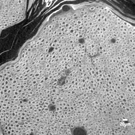
Biological and Polymer Samples
Shown in the example below is a mouse sciatic nerve sample, demonstrating that the system’s ultrahigh resolution clearly details the axons and myelin sheaths
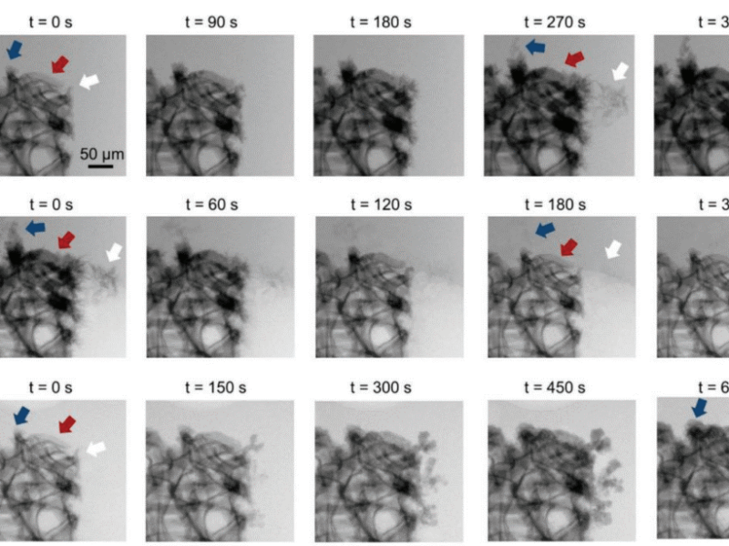
In-situ Microstructural Evolution
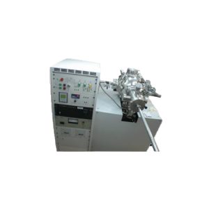
Dendritic growth imaged on PrismaXRM (Sigray’s leading XRM prior to EclipseXRM’s release) on a Zn-ion battery.In-situ cells are manufactured by a variety of manufacturers and can provide heating, T&C, fluid flow, and electrical power
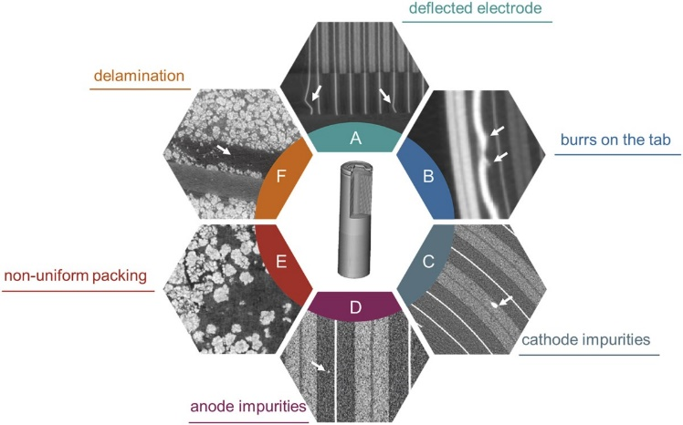
Comprehensive Imaging Solutions for Intact and In-Operando Batteries, and Battery Defects
- Intact Batteries and Batteries in-operando
- Battery defects that can be imaged by Sigray’s x-ray microscope line