Nanotechnology Singapore | Laboratory Equipment | Quasi-S
  • Company
  • Products
  • Services
  • Contact
Nanotechnology Singapore | Laboratory Equipment | Quasi-S
Menu
Hitachi SU7000 Ultra-High-Resolution Schottky Scanning Electron Microscope (SEM)
Click to enlarge
Home Microscopy Electron Microscopy FE-SEM HITACHI SU7000 Ultra-High-Resolution Schottky SEM
HITACHI SU3800/SU3900 SEM with advanced imaging capabilities
HITACHI SU3800/SU3900 SEM
Back to products
Hitachi SU9000 Ultra-High-Resolution Scanning Electron Microscope (SEM)
HITACHI SU9000 Ultrahigh-Resolution SEM

HITACHI SU7000 Ultra-High-Resolution Schottky SEM

The Hitachi SU7000 Ultra-High-Resolution Schottky SEM is designed to meet the diverse needs of modern research and industrial applications.

  • Versatile Imaging Capability: Fast acquisition of multiple signals, from wide field views to sub-nanometer structures.
  • Multi-Channel Imaging: Processes, displays, and saves up to six signals simultaneously for maximum information acquisition.
  • Advanced Observation Techniques: Optimized for large specimens, variable pressure conditions, and in-situ observations.

This advanced SEM provides exceptional imaging performance and user-friendly features, making it an invaluable tool for scientists and engineers.

Add to quote
This product is already in your quote request list.
Browse the list
  • Description
Description

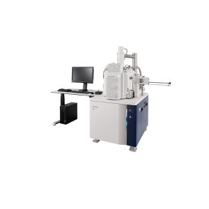
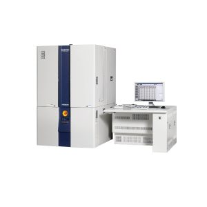
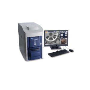
The Hitachi SU7000 Ultrahigh-Resolution Schottky SEM is designed to meet the diverse needs of modern research and industrial applications. This advanced scanning electron microscope offers unparalleled imaging capabilities and user-friendly features.

Features of Hitachi SU7000 Ultrahigh-Resolution Schottky SEM

Versatile Imaging Capability
  • Fast Signal Acquisition: Efficiently acquires multiple signals to address expansive SEM needs, from imaging a wide field of view to visualizing sub-nanometer structures.
  • Advanced Electron Optics: Newly designed electron optics and detection systems allow for the simultaneous acquisition of multiple secondary electron and back-scattered electron signals.
Multi-Channel Imaging
  • Simultaneous Signal Processing: Capable of processing, displaying, and saving up to six signals simultaneously, maximizing information acquisition.
Wide Variety of Observation Techniques
  • Optimized Specimen Chamber and Vacuum System: Designed for large specimen sizes, sample manipulation at various axes, variable pressure conditions, cryogenic conditions, and in-situ observation under heating and cooling.
Microanalysis
  • High-Performance Schottky Emitter: Provides up to 200 nA beam current for various microanalysis applications.
  • Multiple Analytical Options: Supports EDX, WDX, EBSD, cathodoluminescence, and more, enhancing analytical capabilities.
Enhanced Information Acquisition
  • Comprehensive Data Collection: Acquires structural, topographical, compositional, crystallographic, and other types of information with minimal changes to microscope conditions, such as working distance or accelerating voltage.
Single-Scan Multi-Signal Imaging
  • Simultaneous Image Acquisition: Captures surface micro-structural information (UD), surface coating (MD), and overall topographic information (LD) in a single scan.
User-Friendly Interface
  • Intuitive Graphical User Interface: Enhances signal display with a highly flexible screen layout and dual monitor setup.
  • Expandable Observation and Analysis: Features a large specimen chamber and stage, optional camera navigation, various detectors for dynamic observation, and improved PD-BSED response speed.

Ultimately, the Hitachi SU7000 Ultra-High-Resolution Schottky SEM provides exceptional imaging performance and user-friendly features, supporting advanced research and industrial applications. With its versatile imaging capabilities, multi-channel imaging, and comprehensive microanalysis options, this SEM is an invaluable tool for scientists and engineers

Application

Specimen: Fiber with metallic oxide
Detector Selection Under Low-Vacuum Conditions
UVD (SE image)
The oxide dispersion and fiber layering state are observed respectively.

Related Application

Hitachi TM4000II/TM4000PlusII Tabletop Scanning Electron Microscope (SEM)

HITACHI TM4000 II / TM4000 PLUS II Tabletop SEM

Add to quote
This product is already in your quote request list.
Browse the list

OPTIKA Biological Microscopes- Ecovision Series

Add to quote
This product is already in your quote request list.
Browse the list

OPTIKA B-190 Series Biological Microscopes

Add to quote
This product is already in your quote request list.
Browse the list

OPTIKA B-810/ B-1000 Series

Add to quote
This product is already in your quote request list.
Browse the list

OPTIKA Professional Stereo Microscopes – Modular Series

Add to quote
This product is already in your quote request list.
Browse the list

OPTIKA POL Series

Add to quote
This product is already in your quote request list.
Browse the list

OPTIKA FLUO Series

Add to quote
This product is already in your quote request list.
Browse the list

OPTIKA Stereo Microscopes – MX/SFX Series

Add to quote
This product is already in your quote request list.
Browse the list
    © Quasi-S Pte. Ltd. . All right reserved.

    company

    Products

    Services

    contact

    Quasi-S Pte Ltd
    (HQ)

    35 Marsiling Industrial Estate Road 3 #04-03, Singapore 739257.

    T (65) 6383 4386
    F (65) 6383 3071
    E contact@quasi-s.com.sg

    Quasi-S Sdn Bhd
    (Bangi Branch)

    Block Stroma B3-1, UKM-MTDC Smart Technology Centre 43600 Bangi, Selangor, Malaysia

    T (603) 8210 1765
    F (603) 8920 2506
    E inquirybangi@quasi-s.com.my

    Quasi-S Sdn Bhd
    (Penang Branch)

    15-G-26 Bayan Point Medan Kampung Relau 11900 Penang, Malaysia

    T (604) 645 6973
    F (60) 4645 6573
    E inquiry@quasi-s.com.my

    • Company
    • Products
    • Services
    • Contact