")


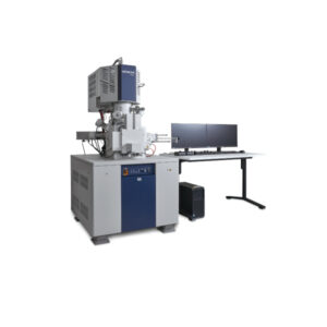
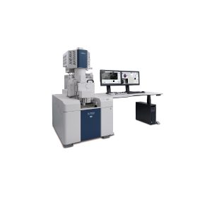
The Hitachi SU8700 Ultrahigh resolution Schottky SEM represents a significant advancement in scanning electron microscopy. This state-of-the-art instrument delivers ultrahigh-resolution imaging and advanced microanalysis, undoubtedly, making it invaluable for researchers and professionals across various disciplines. The Hitachi SU8700 Ultrahigh resolution SEM incorporates a Schottky emitter, providing high probe current and exceptional imaging capabilities, even at low accelerating voltages. Consequently, this feature benefits the examination of beam-sensitive specimens by minimizing damage while maintaining image quality.
Features of Hitachi SU8700 UltraHigh-Resolution Schottky SEM:
Ultrahigh-Resolution and Analytical Capability
- Hitachi’s Schottky emitter delivers ultrahigh-resolution images and ultrafast microanalysis with high probe current.
- Its 0.1kV imaging capability without stage bias indeed expands the application range for beam-sensitive specimens.
- Additionally, users can choose from a multitude of detectors and options to best suit their needs.
Enhanced User Experience with Advanced Automation in Hitachi SU8700 Ultrahigh Resolution Schottky
- The “EM Flow Creator“ software option allows users to configure repeatable SEM operation sequences.
- Users can assemble various SEM functions in the EM Flow Creator’s window by a drag-and-drop method and save them as a recipe for later use.
- Subsequently, once configured, the recipe enables automated data collection under set conditions with high accuracy and repeatability.
Advanced Display and Interface of Hitachi SU8700 Ultrahigh Resolution Schottky
- Dual monitor configuration supports a flexible and highly efficient workspace.
- Users can display and save 6 signals simultaneously to acquire more information in less time.
- Moreover, the system can acquire up to 40,960 x 30,720 pixels of high-resolution information.
Exceptional Field of View and Pixel Resolution
- The large field of view (FOV) combined with high pixel resolution ensures that users can observe even the smallest features of a sample with clarity.
- High-resolution images up to 40,960 x 30,720 pixels ultimately, provide detailed and precise imaging for various applications.
Briefly, the Hitachi SU8700 Ultrahigh resolution SEM excels in automation and user experience. The “EM Flow Creator” software option enables users to configure repeatable SEM operation sequences through a simple drag-and-drop interface. This automation enhances workflow efficiency and ensures high accuracy and repeatability in data collection. Furthermore, the dual monitor configuration supports a flexible and highly efficient workspace, allowing users to display and save multiple signals simultaneously.
In terms of resolution, the Hitachi SU8700 Ultrahigh resolution SEM can acquire high-resolution images up to 40,960 x 30,720 pixels. Admittedly, this level of detail is crucial for applications requiring precise and detailed imaging, such as materials science, nanotechnology, and biological research. Thus, the large field of view (FOV) combined with high pixel resolution ensures that users can observe even the smallest features of a sample with clarity.
Summing up, the Hitachi SU8700 Ultrahigh resolution SEM’s robust construction and innovative technology make it a reliable choice for long-term use. Its advanced features and user-friendly design cater to both novice and experienced users, hence, ensuring that it meets the diverse needs of any laboratory or research facility.
Application

Specimen: Ultrathin section BSE images of Rat Cerebral Cortex
Top-left image was acquired with >120 μm of FOV. The yellow rectangle field in the image is also shown in right image with an increase of digital magnification.
Even after digitally enhancing the original image more than 20 times, the structures of organelle were clearly visible and high quality was maintained.
High-resolution images up to 40,960 x 30,720 pixels are available (*) on SU8700 and SU8600.