Features:
- Versatile Imaging Capability
– Fast acquisition of multiple signals to address expansive SEM needs, from imaging a wide field of view to visualizing sub-nanometer structures and everything in between
– Newly designed electron optics and detection systems, allows for efficient simultaneous acquisition of multiple secondary electron and back-scattered electron signals - Multi-Channel Imaging
– Capable of processing, displaying, and saving up to 6 signals simultaneously to maximize information acquisition - Wide Variety of Observation Techniques
– Specimen chamber and the vacuum system are optimized for:
・Large specimen size
・Sample manipulation at various axes
・Variable pressure conditions
・Cryogenic conditions
・Heating and cooling in-situ observation - Microanalysis
– Schottky emitter electron gun, provides up to 200 nA beam current, for various microanalysis applications
– Design to incorporate multiple analytical options including EDX, WDX, EBSD, cathodoluminescence, and more - Enhanced Information Acquisition
- Acquisition of structural, topographical, compositional, crystallographic, and other types of information, by minimizing changes to microscope conditions, such as working distance or accelerating voltage
- Single-Scan Multi-Signal Imaging
- Simultaneous image acquisition for surface micro-structural information (UD), surface coating (MD), and overall topographic information (LD)
- Intuitive graphical user interface
- Enhance signal display
- Highly flexible screen layout
- Dual monitor
- Expandable observation and analysis
- Large specimen chamber and large stage
– Optional Camera Navigation
– Various detectors enabling dynamic observation
– Detector selection under low vacuum conditions
– Improved PD-BSED response speed
Application
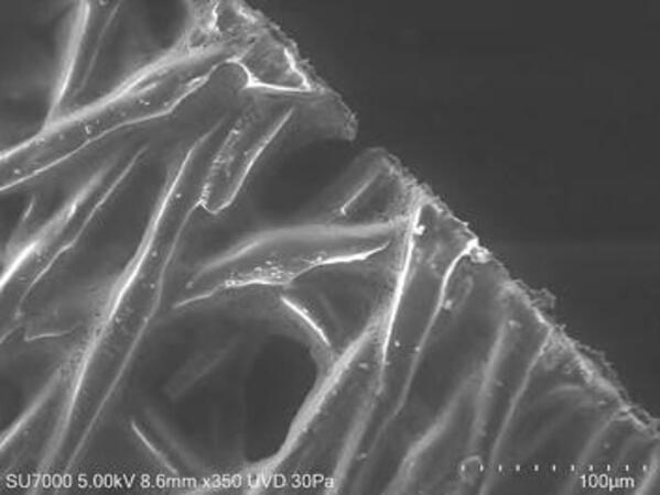
Specimen: Fiber with metallic oxide
Detector Selection Under Low-Vacuum Conditions
UVD (SE image)
The oxide dispersion and fiber layering state are observed respectively.